Best Practices When Selecting Bone Samples
Bones and teeth can be used for a variety of different isotopic analyses, including Oxygen (δ18O), Strontium (87Sr/86Sr), Lead (Pb), Carbon (δ13C), Nitrogen (δ15N) and dating (radiocarbon and in some cases Uranium-Thorium). They are particularly useful as they tend to have high preservation potential and are therefore quite common in fossil and archaeological records. Regardless of the isotopic analysis in question, there are important requirements for bone samples to ensure they produce accurate results.

Following burial, bone diagenesis occurs – a process wherein the physical and chemical bone characteristics are altered after burial. Understanding the process of diagenesis is vital as the elemental and isotopic compositions of bones and teeth are influenced by a variety of environmental and lifestyle factors – including diet, water uptake, local geology and movement through an individual’s lifetime. However, the reliable application of isotopic methods on bone samples depends on the assumption that the chemical composition remains the same after death and burial. If any chemical alteration occurred thereafter, this could negatively impact the certainty and reliability of isotopic measurements and interpretation of results. Vitally for bones and teeth, the inorganic (e.g. phosphate, carbonate) portion tends to be better preserved than the organic portion (collagen) – making inorganic structures of bones/teeth more optimal than inorganic structures for isotopic analyses.
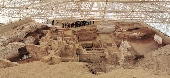
 The Çatalhöyük settlement in Anatolia, where the community buried their dead within the village beneath the buildings.
Burial conditions
Bones tend to retain their chemical properties best when they are deposited and buried in relatively cool, dry conditions with limited water throughflow, anoxic, low pH conditions – resulting in a smooth surface, high structural integrity surface. These bones tend to be quite sheltered, not exposed to regular seasonality including freeze-thaw cycles; such environments tend to occur in areas with low porosity soils often below the water table where water and oxygen flow are restricted. Conversely, bones buried in relatively warm, humid environments – particularly when combined with acidic, porous soils and/or exposure to sun, wind or seasonal cycles – tend to have a cracked exterior, with low structural integrity throughout resulting in a high probability of chemical alteration. Specifically, the collagen within the bone structure tends to leach out very quickly when the bone comes into contact with water. These bones tend to be less sheltered, coming into contact with groundwater or other moist conditions – sometimes resulting in micro-bacterial attack and degradation through the bone surface.
Depending on the local environmental conditions and degree of environmentally-driven diagenesis, the interior bone sections may be less susceptible to chemical alteration compared to the exterior and thus may be still usable for isotopic analyses. Tooth samples tend to fare better in harsh environments as the enamel coating helps preserve underlying tooth proteins. Regardless of the type of bone – when the structure is compromised, it’s best to submit larger bone samples to ensure the outer layer can be removed prior to analysis. Elemental maps of bone cross sections have demonstrated variations in elemental concentrations (e.g. Rasmussen et al. 2019), which can be useful in selecting the optimal samples for further analysis prior to submission for dating and other isotopic analyses.
Pre-burial burning & charring
Beyond the local burial environment, any bone alteration that occurred before burial can impact the reliability of the isotopic signatures – particularly burning and charring which often occurred for cooking and ceremonial purposes. The quality of the bone sample depends on the level of heat applied to the bone as well as the oxygen availability in the environment during burning. When bones are burned at relatively low temperatures (<500°C), they tend to have fragile structures due to higher porosity than those burned at higher temperatures (>500°C) (Marques et al. 2021a). Whilst higher temperatures are better for structural preservation, the inorganic portion of bones often disappear quickly at relatively low temperatures (≈300-450°C) – leaving only the organic portion for analysis. However, this process occurs slower in anaerobic environments. The burning severity may be directly connected to the location of the bone within a fire – Stiner et al. (1995) demonstrated that bones burned within an open fire exhibited more extensive burning than those burned within the depths of coal beds.
Whilst it can be difficult to predict the level of burning that occurred, in cases where the bones were burned in an aerobic environment, the color of the bone can provide clues to the level of burning. Marques et al. (2021b) demonstrated a clear gradient of bone color from black (400°C) at relatively low temperatures to brown-gray at moderate temperatures (600-700°C) and eventually white at very high temperatures (900-1000°C) after advanced burning; no gradient was found between the same temperature classes in anaerobic conditions. However, in low temperature / light burns the bones may remain lightly shaded before turning black or demonstrate localized burns along their structure (Stiner et al. 1995).
Fossilization
In cases where a human or animal is buried by sediment soon after death with limited exposure to the environment and scavengers, the process of fossilization may occur. In such cases, the organic portions of the bone slowly breakdown, leaving the hard inorganic material behind with a porous structure. The bone pores eventually get infilled by surrounding minerals (e.g. calcium carbonate), precipitating into the pore spaces – resulting in a bone that exhibits a rock-like structure. When analyzing such samples, it’s important to only analyze the segments of the original inorganic bone material rather than the fossilized infill. In order to differentiate between the two materials, XRD (X-Ray Diffraction) can be undertaken. For example, Storm et al. (2013) identified areas of original bone material using XRD laser tracks within the bone structure in order to pinpoint subsampling areas for Uranium-series “spot” dating.
Submission
When submitting your bone samples for isotope analysis or dating, it is important to note the burial conditions and any burning processes that occurred. Isobar Science presently accepts bone samples for Lead (Pb), Uranium-Thorium (U-Th) dating, and Strontium (87Sr/86Sr) analyses.
| Bone Type | Lead (Pb) | U-Th Dating | Strontium (87Sr/86Sr) |
| Non-ashed | 200mg | 100-150mg | 500mg |
| Ashed | 500mg | Not suitable | 200mg |
| Burnt | N/A | Not suitable | 500mg |
| Fossilized | 500mg** | 100-150mg* | 500mg** |
*Only accepted if XRD complete
**Analysis can be conducted but the results may be affected by post deposition processes
References
Marques, M.P.M., Batista de Carvalho, L.A.E., Gonçalves, D., Cunha, E. and Parker, S.F., 2021a. The impact of moderate heating on human bones: An infrared and neutron spectroscopy study. Royal Society Open Science, 8(10), p.210774.
Marques, M.P.M., Gonçalves, D., Mamede, A.P., Coutinho, T., Cunha, E., Kockelmann, W., Parker, S.F. and Batista de Carvalho, L.A.E., 2021b. Profiling of human burned bones: oxidising versus reducing conditions. Scientific Reports, 11(1), pp.1-13.
Rasmussen, K.L., Milner, G., Skytte, L., Lynnerup, N., Thomsen, J.L. and Boldsen, J.L., 2019. Mapping diagenesis in archaeological human bones. Heritage Science, 7(1), pp.1-24.
Stiner, M.C., Kuhn, S.L., Weiner, S. and Bar-Yosef, O., 1995. Differential burning, recrystallization, and fragmentation of archaeological bone. Journal of archaeological science, 22(2), pp.223-237.
Storm, P., Wood, R., Stringer, C., Bartsiokas, A., de Vos, J., Aubert, M., Kinsley, L. and Grün, R., 2013. U-series and radiocarbon analyses of human and faunal remains from Wajak, Indonesia. Journal of human evolution, 64(5), pp.356-365.
Image References
1. https://www.pexels.com/photo/dinosaur-and-human-skeletons-12227297/
2. https://en.wikipedia.org/wiki/File:Çatalhöyük,_7400_BC,_Konya,_Turkey_-_UNESCO_World_Heritage_Site,_08.jpg
3. Storm et al. (2013)
